
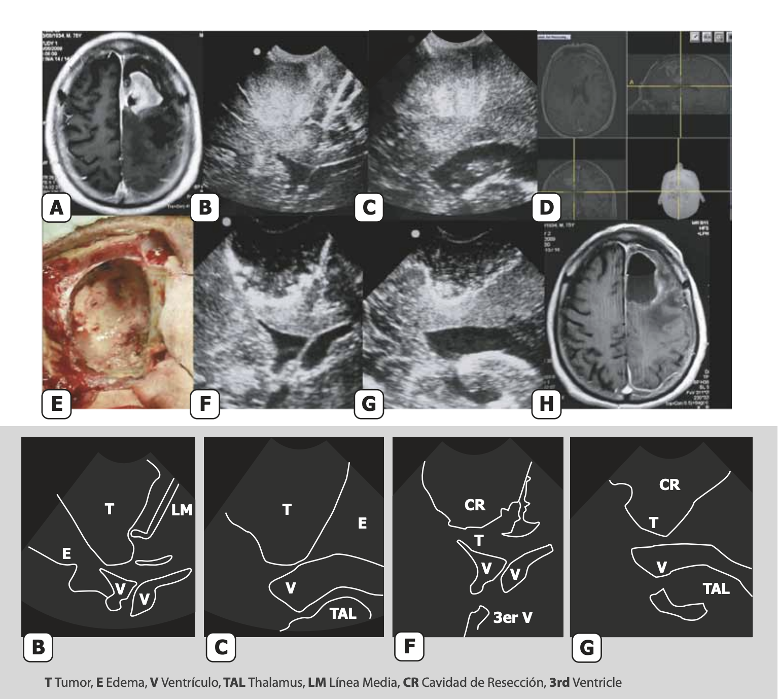
Completed in 2010 after more than 15 years of clinical and surgical work, Dr. Eduardo Lovo's Atlas of Intraoperative Ultrasound in Neurosurgery was created to address a concrete challenge: the visual learning curve every neurosurgeon faces when integrating ultrasound into the operating room.
What the atlas covers
The atlas combines fundamental technical principles with real operative cases, establishing direct correlations between intraoperative ultrasound images and MRI. That visual parallel is its core contribution: it allows the surgeon to learn ultrasound "language" using the MRI reference they already know.
Although technology has evolved since publication, the anatomical principles, image patterns, and practical concepts presented here remain remarkably relevant today.
Dedicated atlases of intraoperative neurosurgical ultrasound remain uncommon. This is likely one of the few comprehensive examples produced in Spanish.
Open access
Dr. Lovo is making the complete atlas freely available—at no cost—particularly for residents, young neurosurgeons, and programs beginning their learning curve in intraoperative ultrasound.