
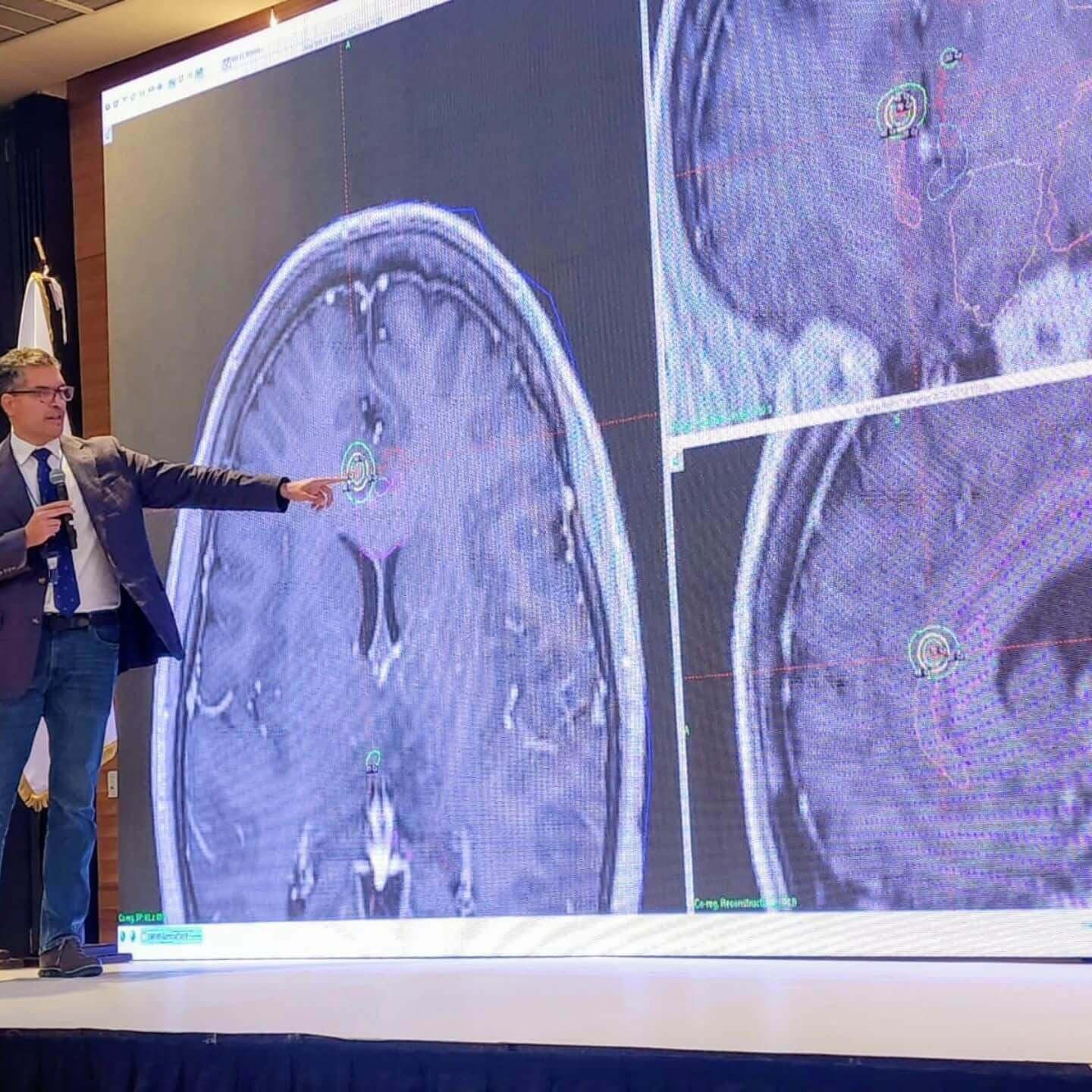
For most of my professional life, I have worked inside a paradox. A neurosurgeon must be willing to enter the most complex organ in the human body while developing an increasingly profound respect for what should not be touched.
That paradox has shaped more than two decades of my work in brain tumor surgery, image-guided neurosurgery, awake craniotomy, functional neurosurgery and stereotactic radiosurgery in El Salvador and Central America. The technologies have changed dramatically during that time. The fundamental problem has not.
When we operate on the brain, we are not simply treating tissue. We are operating beside language, movement, memory, emotion and the biological architecture from which a human being experiences the world.
Learning to see beyond the surgical field
My early experience with image-guided neurosurgery began during my neurosurgical training at the Pontificia Universidad Católica de Chile. Intraoperative ultrasound offered something extremely valuable: real-time anatomical information during surgery.
After returning to San Salvador, El Salvador, we incorporated intraoperative ultrasound into the neuro-oncology program we were developing. By 2007 it had become part of our image-guided approach to brain surgery, and in 2010 we completed an atlas dedicated to intraoperative ultrasonography of the central nervous system.
Its purpose was practical. We placed ultrasound images obtained during surgery alongside corresponding MRI images so that neurosurgeons could learn to translate one visual language into another.
Today this may seem straightforward. At the time, it represented an important change in how we approached brain tumor surgery. The surgeon no longer depended exclusively on what could be seen through the microscope or on an MRI obtained before entering the operating room. We could obtain new information while the operation was actually occurring.
For our program, this was one of the foundations of image-guided neurosurgery.
The brain moves. The map must move with it.
Preoperative MRI provides an extraordinary map, but the brain is not static during surgery. After the skull and dura are opened, cerebrospinal fluid is released and tumor resection begins, anatomical structures can shift. The relationships represented on a preoperative scan may no longer correspond perfectly with the anatomy in front of the surgeon.
Neuronavigation gives us a map. Intraoperative ultrasound helps us update it.
But over time we learned that even a perfect anatomical map would not be enough, because anatomy does not tell the entire story of the brain.
From image guidance to awake brain surgery
A tumor can have a visible border on MRI. Language does not. Movement does not. Memory does not.
Functional networks may pass through, around or immediately beside something that appears clearly defined on an image. This realization made awake craniotomy and functional mapping increasingly important components of our brain tumor program.
Our awake-craniotomy experience began in 2007. A published analysis of our experience through 2018 described 218 attempted awake craniotomies, of which 213 were successfully completed. Since that published series, the accumulated clinical experience of our program has grown to more than 500 awake craniotomies.
That distinction matters. The earlier number represents the published cohort; more than 500 represents the subsequent cumulative clinical experience of the program.
Over time, awake surgery became more than an extraordinary operation reserved only for tumors immediately adjacent to language cortex. For selected patients, it developed into a reproducible approach to supratentorial brain tumor surgery.
Sometimes cortical or subcortical mapping is required. Sometimes it is not. The value of awake surgery extends beyond mapping alone. It can allow continuous neurological assessment, early recognition of functional change and direct interaction with the person whose brain we are operating on.
And that last point is more important than it may initially appear.
Precision is not presence
Neurosurgery teaches us to respect precision. Millimeters matter. A few millimeters can separate tumor from motor pathways, a radiosurgical target from an optic structure, or an effective intervention from permanent neurological injury.
But years of brain surgery have taught me something else: precision is not presence.
A navigation system can tell me where I am. A tractography reconstruction can suggest where fibers travel. An ultrasound image may tell me whether tumor remains.
But none of them can fully tell me what is happening when the person lying on the operating table suddenly hesitates before naming an object, loses a word that was available seconds earlier, or senses that something inside his own experience has changed.
Silence can become information. Hesitation can become information. A pause can contain meaning. There are moments when the surgeon must become more than a technician.
Brain tumor surgery is ultimately surgery of preservation
Modern neurosurgery frequently speaks of maximal safe resection. It is an important principle, but I believe the order of the words matters.
The objective should not be maximal removal followed by an attempt to remain safe. The objective is the most appropriate resection while preserving what allows the patient to return to a life he or she recognizes as their own.
The best brain tumor operation is therefore not necessarily the operation that removes the greatest volume of tissue. It is the operation that most precisely understands the boundary between what can be removed and what must be preserved.
Image guidance helps us define that boundary anatomically. Awake surgery helps us define it functionally. Microsurgical technique allows us to respect it physically. Radiosurgery offers another strategy when crossing that boundary surgically would impose too high a neurological price.
These technologies are not competitors. They are different instruments serving the same patient.
Building advanced brain tumor surgery in Central America
Much of modern neurosurgical technology was developed in major centers in North America, Europe and Asia. Our experience was built in Central America.
That has shaped the way I understand technology. Sophisticated medicine cannot consist simply of acquiring sophisticated machines. It requires identifying which technologies solve meaningful clinical problems, integrating them into reproducible clinical systems and making them work within the economic and social reality in which patients actually live.
Image-guided surgery, intraoperative ultrasound, awake craniotomy, cortical and subcortical mapping, stereotactic radiosurgery and modern neuro-oncology did not develop in our program as isolated acquisitions. They became parts of an architecture.
Over time, that architecture helped us construct a brain tumor program in El Salvador capable of approaching increasingly complex disease while preserving a simple principle: technology must serve the patient; it must never become the purpose of the operation.
Our experience in Central America has also reinforced another conviction. Geography should influence how we solve a medical problem. It should not determine the quality of medicine a patient deserves.
From structures to networks
My own understanding of the brain also changed during these years. At first, we learn structures. Then pathways. Eventually, networks.
Language, movement, pain, memory, emotion and behavior cannot always be understood as isolated anatomical territories. They arise from distributed systems that interact continuously.
That transition has increasingly influenced my work in functional neurosurgery and stereotactic radiosurgery. Radiosurgery historically gave us the ability to deliver highly focused energy to a precisely defined anatomical target.
But when we begin thinking in networks, a different possibility emerges: in some disorders, perhaps the objective is not simply to destroy a structure but to modify the behavior of a pathological circuit.
That concept has helped guide our work toward increasingly individualized and connectivity-informed approaches to pain and other functional disorders.
And with that capability comes a much larger question: if we can identify the networks involved in movement, pain, emotion or behavior, how far should we intervene?
When the surgical question becomes a human question
That question eventually became larger than neurosurgery.
For years, I entered the brain as a surgeon. I removed tumors. I stimulated cortex. I interrupted pathological circuits. I delivered focused radiation to structures only millimeters in diameter.
But the more technically capable we became, the more another question began to matter: What exactly are we touching?
Not anatomically. Humanly.
A brain is tissue. But somewhere within that tissue emerge memory, fear, desire, language, imagination and the experience of being a person.
These questions have increasingly become part of my writing beyond the operating room, as I explore the intersection of neuroscience, technology, identity and the ethical responsibilities created by our growing ability to read and modify the human brain.
My career makes an argument against technology impossible. Technology has allowed us to reach tumors once considered inaccessible, preserve functions we once sacrificed, treat disease without opening the skull and return patients to lives they might otherwise have lost.
But technological capability creates responsibility.
The question is no longer only: What can we do to the brain?
It is also: What should we do once we can?
Neurosurgery in the age of artificial intelligence
We are entering another transition. Artificial intelligence is increasingly capable of interpreting medical images, integrating large quantities of clinical information and recognizing patterns that may be difficult for unaided human perception to identify.
In neurosurgery, the potential convergence is extraordinary: MRI, tractography, intraoperative ultrasound, neuronavigation, physiological monitoring, functional mapping, genomics and clinical outcomes may increasingly be integrated around a single patient.
The operating room of the future may understand anatomy, connectivity and probability at a level no individual surgeon could reproduce alone. That is enormously promising.
But intelligence without empathy is an incomplete instrument.
The future of neurosurgery cannot simply become a more computationally powerful version of its past. Technology must remain subordinate to the human being whose brain generated the data in the first place.
The important question is not only what the machine can do. It is whom it serves.
More than 500 awake craniotomies later
When I look today at the intraoperative ultrasound atlas we completed more than fifteen years ago, I see an early chapter of this same journey.
First we learned to see. Then to navigate. Then to map. Then to listen.
And progressively, to understand the brain not simply as an organ divided into anatomical territories, but as an interconnected system from which human function and experience emerge.
More than 500 awake craniotomies later, I remain more impressed by the brain than when I began. But I am also more cautious.
Experience does not necessarily produce certainty. Often it produces a deeper understanding of uncertainty.
Perhaps one of the deepest lessons of brain surgery is therefore restraint.
A technically competent neurosurgeon must know how to enter the brain. A mature surgeon must also know when to stop.
And that principle may ultimately extend far beyond the operating room.
As our capacity to read, predict and modify the human brain continues to grow, the measure of our progress will not simply be how deeply we learn to enter the human mind.
It may be whether we develop the wisdom to know when to take our hands away.