
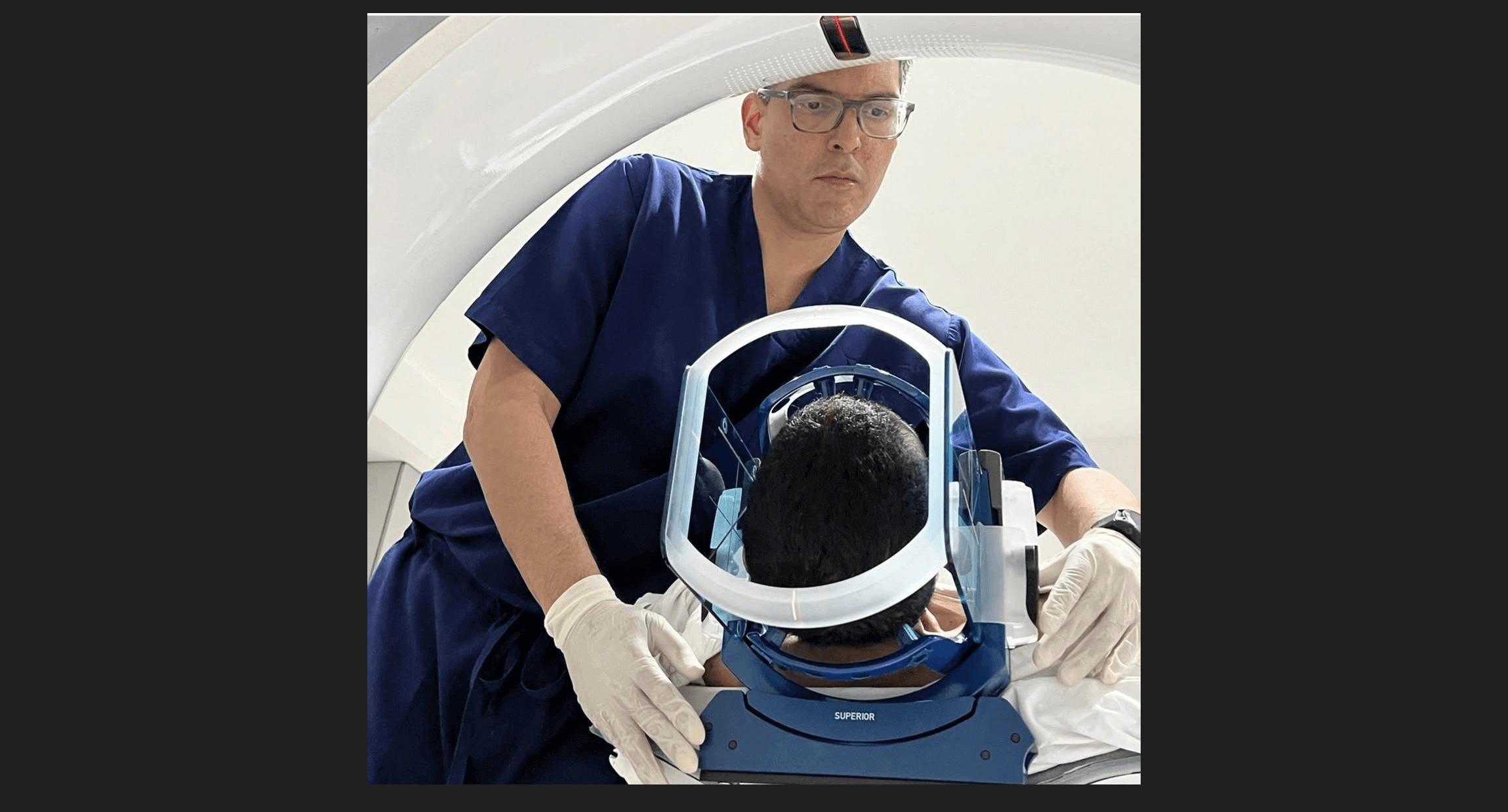
More than fifteen years ago, we created an atlas of intraoperative ultrasound in neurosurgery in El Salvador. Looking at it today, I see much more than an old educational project. I see one of the early foundations of the brain tumor surgery program we were beginning to build.
The atlas, completed in 2010, was designed to solve a very practical problem: how does a neurosurgeon translate the familiar anatomy of a preoperative MRI into the very different image seen on an ultrasound screen during an operation?
That question became part of a much larger one: how can imaging, functional mapping and surgical judgment be integrated to remove a brain tumor while preserving as much normal brain function as possible?
For our program, that was the beginning of image-guided neurosurgery.
From MRI to the operating room
Brain tumor surgery is fundamentally a problem of anatomy in motion.
MRI provides an extraordinary map before surgery. But once the skull and dura are opened, cerebrospinal fluid is released and tumor resection begins, the brain can shift. The anatomical relationships seen on the preoperative scan may no longer be exactly the same.
Intraoperative ultrasound provided us with something fundamentally different: real-time information from inside the operation.
Our early experience with this technology began during my neurosurgical training in Chile and was later incorporated into practice in El Salvador. By 2007, dedicated intraoperative ultrasound was being used in our neuro-oncology program, and by early 2010 it had already been applied in more than 85 operations involving tumors, vascular malformations, abscesses, epilepsy surgery and hematomas.
This work was not isolated from our academic development. We had already published on the technical aspects of intraoperative ultrasound in neurosurgery in 2006, followed by a report describing the evolution of tumor neurosurgery in El Salvador in 2009.
The atlas completed in 2010 brought many of those lessons together visually. Its central idea was simple: pair intraoperative ultrasound with corresponding MRI images so that young neurosurgeons could gradually learn to recognize the anatomy of the brain through a new imaging language. The work included technical principles and case-based examples involving temporal lesions, high- and low-grade tumors, posterior fossa lesions and hematomas.
Image guidance alone is not enough
A tumor can be visible on an MRI and still be intimately connected with the structures that make a person who he or she is.
Language, movement, sensation, memory and other functions do not respect the borders of a tumor.
For tumors close to eloquent brain regions, knowing where the tumor is is only part of the problem. The surgeon also needs to know what the surrounding brain is doing.
This is where awake craniotomy and cortical mapping became another major component of our program.
During an awake craniotomy, selected patients can perform language, motor or cognitive tasks while specific cortical and subcortical areas are tested during surgery. The objective is not simply to remove the largest possible amount of tumor. It is to pursue the greatest appropriate resection while respecting the functional boundaries of the individual patient.
That distinction is central to modern brain tumor surgery.
Our group began reporting its experience with awake craniotomy more than a decade ago. A later published analysis reviewed procedures performed between January 2007 and July 2018. In that series, 218 awake craniotomies were attempted and 213—98.1%—were successfully completed. Eighty percent of the patients had tumors, and the series included lesions located in eloquent, near-eloquent and non-eloquent regions.
The publication concluded that awake craniotomy was safe and well tolerated in the majority of patients and was useful for tumor removal in eloquent cortex.
Since that published series, our cumulative clinical experience has continued to grow and has now exceeded 500 awake craniotomies.
That current figure represents our institutional clinical experience rather than the cohort reported in the 2018 publication, and the distinction is important: published data should remain identifiable as published data, while later accumulated experience should be described as such.
When imaging and function come together
Perhaps the most important development was not any individual technology but the integration of several of them.
In a 2012 publication, our group reported patients with gliomas undergoing surgery using awake craniotomy, image-guided surgery, intraoperative ultrasound and intraoperative MRI. In that initial series, ultrasound identified the extent of tumor resection with an 89% reliability compared with high-field intraoperative MRI, and complete radiological resection was achieved in the reported cases.
That experience reflects the philosophy that ultimately shaped our brain tumor program.
Neuronavigation provides a map.
Ultrasound updates that map in real time.
Awake mapping identifies the functional boundaries that imaging cannot see.
Microsurgical technique determines what can be removed safely.
And radiosurgery provides another option when surgical removal is not the best answer for the entire lesion.
Excellence in brain tumor surgery is therefore not defined by a single machine or a single operation. It comes from integrating anatomy, imaging, function, technology and judgment around the individual patient.
A distinctive experience developed in Central America
Much of the literature describing advanced image-guided and awake brain tumor surgery historically comes from major centers in North America, Europe and Asia.
Our experience developed in San Salvador, El Salvador.
That matters.
The 2009 publication Avances en neurocirugía tumoral: El Salvador, C.A. documented the development of these techniques locally, including stereotactic and frameless image guidance, intraoperative ultrasound, awake surgery and cortical mapping.
The subsequent publications on intraoperative ultrasound, multimodality imaging and awake craniotomy created a verifiable academic record of how a sophisticated neuro-oncology surgical program was being constructed in Central America.
This does not mean that advanced brain tumor surgery belongs to one institution or one surgeon. Quite the opposite. It demonstrates that the principles of modern neuro-oncology surgery can be developed, adapted and sustained outside the traditional centers where these technologies originated.
For me, that may be one of the most important lessons of the last two decades.
Geography should influence how we solve a problem. It should not determine the level of medicine a patient deserves.
Why intraoperative ultrasound still matters in the age of AI
There is another reason I believe this old atlas deserves to be made available again.
Intraoperative ultrasound is becoming newly relevant.
Artificial intelligence, computer vision and advanced image registration are increasingly being applied to intraoperative ultrasound. Recent research has demonstrated AI systems capable of detecting brain tumors in ultrasound images in real time, while other work is exploring the transformation of intraoperative ultrasound into MRI-like representations that may make the images easier to interpret and integrate with existing surgical platforms.
The fundamental challenge, however, is the same one we were trying to address more than fifteen years ago:
How do we understand what we are seeing inside the brain while the operation is actually happening?
The machines are changing.
The computational tools are changing.
The anatomy is not.
That is why many of the principles contained in this atlas remain surprisingly recognizable today.
Making the atlas available again
I had believed for some time that this work had been lost.
Finding it again more than fifteen years later felt like recovering a piece of the history of our program.
Dedicated atlases of intraoperative ultrasound in neurosurgery remain relatively uncommon, and extensive resources of this type in Spanish are particularly unusual. I have therefore decided to make the atlas freely available for educational purposes, especially for neurosurgical residents, young neurosurgeons and programs beginning their learning curve in intraoperative ultrasound.
The Spanish edition will be available through this website, and an English version will also be made available shortly.
What began as a collection of ultrasound images became part of something much larger: the development of image-guided neurosurgery, awake brain mapping and a mature brain tumor program in Central America.
More than fifteen years later, after more than 500 awake craniotomies and continued evolution in navigation, radiosurgery and functional brain mapping, I still believe in the same principle that motivated that early work:
The best brain tumor surgery is not simply the surgery that removes the most tumor. It is the surgery that understands, with the greatest possible precision, what can be removed—and what must be preserved.